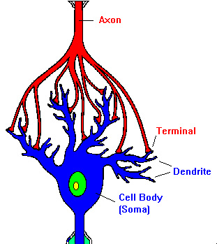

Somatic Golgi and GOPs are considered (central) stations of the secretory pathway and MT nucleation sites. Both single- and multi-compartment GOPs are present in Drosophila neurons, and most of them are located in the dendritic shaft and branching points, respectively.

In contrast to vertebrate GOPs, Drosophila GOPs are widespread in the dendritic arbors including distal dendrites, and are enriched at branch points. In Drosophila (da sensory neurons), the somatic Golgi apparatus contains cis-, medial- and trans-Golgi components as in vertebrate neurons, but lacks ribbon structure instead, Golgi forms mini-stacks or “ring”-like stacks. Smaller Golgi structures that lack many protein components for sorting and organization of Golgi cisternae are named Golgi satellites, which were identified in distal dendritic arbors in cultured rat neurons. Multi-compartment GOPs are largely restricted to one primary dendrite and are often found in the proximal region. One potential target for therapeutic interventions is brainderived neurotrophic factor (BDNF), known to promote neuronal growth and survival and decrease in aging (Binder & Scharfman, et al ). In mice (hippocampal neurons), somatic Golgi shows a ribbon structure and appears to face and extend into primary dendrite(s). Developing approaches aimed at promoting neurogenesis and/or dendritic morphology is necessary. Golgi apparatus in vertebrate and Drosophila neurons. on behalf of KeAi Communications Co., Ltd. We describe the organization of the Golgi apparatus in neurons, review the current understanding of Golgi function in dendritic morphogenesis, and discuss the current challenges and future directions.ĭendrite Golgi Golgi outposts Microtubule Neurodevelopmental disorders Secretory pathway. However, the organization and function of Golgi in dendrite development and its impact on neurological disorders is just emerging and so far lacks a systematic summary. The neuronal Golgi apparatus shares common features with Golgi in other eukaryotic cell types but also forms distinct structures known as Golgi outposts that specifically localize in dendrites. The Golgi apparatus occupies the center of the secretory pathway and is regulating posttranslational modifications, sorting, transport, and signal transduction, as well as acting as a non-centrosomal microtubule organization center. While dendritic morphology ranges from relatively simple to extremely complex for a specified neuron, either requires a functional secretory pathway to continually replenish proteins and lipids to meet dendritic growth demands. Their development is tightly controlled and abnormal dendrite morphogenesis is strongly linked to neurological disorders. Dendrites are specialized neuronal compartments that sense, integrate and transfer information in the neural network.


 0 kommentar(er)
